What Is A Hemangioma Of T9 Vertebrae
T11 and T12 vertebrae are particularly flexible sections of the spine and are subsequently the most common areas of the thoracic spine to get damaged. Spinal cord injuries in the T9 - T12 vertebrae often result from severe trauma or compression fractures bone damage that leads to a shrinkage of the vertebrae.
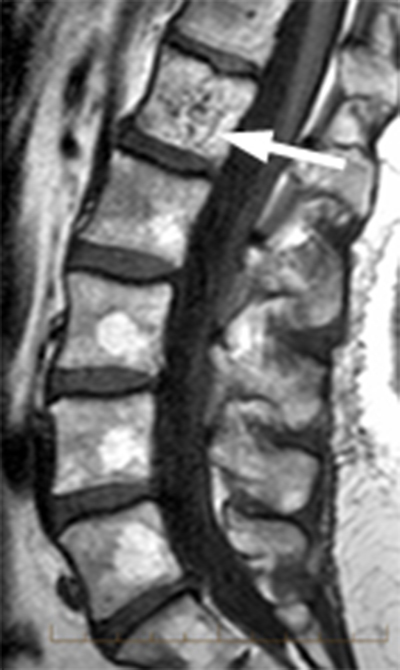
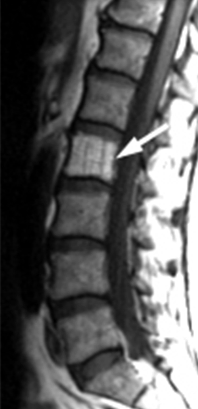
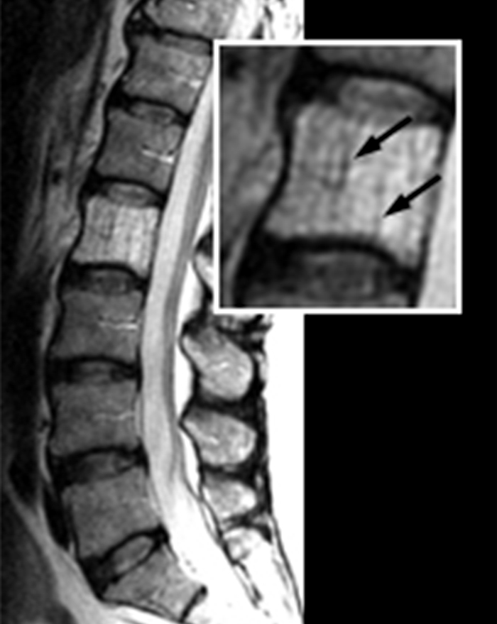 Vertebral Hemangioma Radsource
Most cases show no symptoms.
What is a hemangioma of t9 vertebrae. Also referred to as vertebral hemangioma it is non-cancerous and has few symptoms. Hemangioma is the most common benign vascular tumor of bone. A spinal hemangioma is a tumor that mostly occurs in the thoracic and the lumbar spine.
They are present in about 10 of people. The majority of these tumors will be discovered only incidentally in passing or not at all will never cause symptoms. Most vertebral hemangiomas are located in either the thoracic upper- to mid- or lumbar lower spine.
The term hemangioma refers to a mass of blood vessels that commonly occur on the subcutaneous tissues. What Is a Hemangioma. This is why most people do not realize that they have this condition unless an imaging test for another condition reveals its presence.
These growths near the spine are not as dangerous as malignant spinal tumors but they should be treated anyway as they can seriously interfere with the nerves and other structures in the spine. The typical location of the hemangioma in the spine. In fact they are the most common benign spinal tumors.
Vertebral hemangioma VH is virtually vascular malformation which is usually asymptomatic. Hemangioma is a vascular tumor which is a tangle of interwoven and modified vessels. A hemangioma can occur anywhere on the body but most commonly appears on the face scalp chest or back.
These growths classically appear in the thoracic and lumbar spine located in the mid to lower back. Aggressive vertebral hemangiomata are a rare form of vertebral hemangiomata where significant vertebral expansion extra-osseous component with epidural extension disturbance of blood flow and occasionally compression fractures can be present causing spinal cord andor nerve root compression 12. Although rare displacement of the T9 vertebra may cause severe symptoms in the kidney area as the.
Five days later Im still awaiting results on that. Vertebral hemangiomas are a common etiology estimated to be found in 10-12 of humans at autopsy. While the T2-T9 vertebrae are often linked descriptively the T9 vertebra has certain particularities.
Spinal hemangioma also called vertebral hemangioma is a benign tumor that develops from blood vessels in the bones of the spine or vertebrae. A spinal hemangioma is a benign vascular tumor of the spine. Only 37 of VH may become active and symptomatic and 1 may invade the spinal canal andor paravertebral space.
Commonly these are benign lesions that are found incidentally during radiology studies for other indications. A spinal hemangioma or a hemangioma in spine is a benign tumor that may develop in the bony segments of the spinal column. In the human body there are all in all 33 bones in the spine which are also known as vertebrae.
They are benign in nature and frequently asymptomatic. However involvement of long and short tubular bones has been reported. While the tumor is not dangerous it can cause pain and discomfort and treatment may be recommended for these reasons.
The T9 vertebra is one of the lowest positioned of the twelve 12 thoracic vertebrae and thus one of the largest. Thoracic hemangioma is a big medical term for a benign spinal tumor. Hemangiomas most often appear in adults between the ages of 30 and 50.
Treatment protocols for active or aggressive VHs are still in controversy. Flawed vessels of the vertebra is formed inside the tumor. This type of tumor is most frequently diagnosed in patients between the ages of 30 and 50 and may not cause noticeable symptoms according to Scoliosis and Spine Associates.
The radiologist couldnt confirm that it was a hemangioma so they recommended a CT scan. Usually there is damage to the vertebral bodies but the possibility of tumor growth and cartilage layers. I then got a thoracic MRI which showed a lesion in the T9 vertebral body.
Spinal hemangiomas are widespread. Reported treatments include radiotherapy vertebroplasty direct alcohol injection embolization surgery and a combination of these modalities. For that reason its especially important that your doctor know your complete medical history and perform both general physical and neurological exams.
They are very common and occur in approximately 10 percent of the worlds population. Only rarely are they found in the cervical spine neck. A lumbar hemangioma is a benign blood vessel tumor that grows along one or more vertebra of the lower back.
Spinal hemangiomas usually develop in the bones of the spine known as vertebrae. It looks like a rubbery bump and is made up of extra blood vessels in the skin. A hemangioma he-man-jee-O-muh is a bright red birthmark that shows up at birth or in the first or second week of life.
Symptoms if they do occur are usually related to large hemangiomas trauma the hormonal and hemodynamic changes of pregnancy or osseous expansion and extra-osseous. Most hemangiomas involve the vertebral body or skull and involvement of other bones is rare. Vertebral tumors sometimes may be overlooked because their symptoms resemble those of more-common conditions.
Spinal hemangiomas are benign tumors that are most commonly seen in the mid-back thoracic and lower back lumbar. The ninth thoracic vertebra T9 communicates directly with the adrenal glands via nerves. Given below is some information on what causes an atypical hemangioma in spine and how it can be treated.
A vertebral hemangioma is a vascular lesion within a vertebral body. Instead of two costal demi-facets superior and inferior on the exterior of the centrum it typically has a single superior costal demi-facet above which together with facets on the Continue Scrolling To Read More Below.
 Vertebral Hemangioma Radsource
 Vertebral Hemangioma Radsource
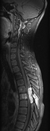
 Symptomatic Thoracic Vertebral Hemangioma A Case Report And Literature Review1 Archives Of Physical Medicine And Rehabilitation
 Vertebral Hemangioma Radsource
 Aggressive Vertebral Hemangioma Radiology Reference Article Radiopaedia Org
Vertebral Hemangioma Radsource
 Hemangioma Vertebral Youtube
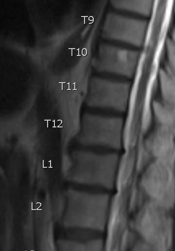
 16 15 Hemangioma Of The T9 Vertebral Body In An 81 Year Old Woman Download Scientific Diagram
A 49 Year Old Female Patient Was Diagnosed As Having T9 Whole Vertebral Download Scientific Diagram
 16 15 Hemangioma Of The T9 Vertebral Body In An 81 Year Old Woman Download Scientific Diagram
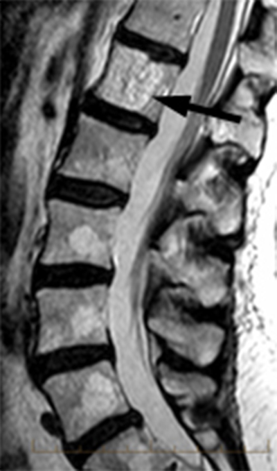
 Aggressive Vertebral Hemangioma Involving T8 Vertebra In A 58 Year Old Download Scientific Diagram
 Symptomatic Thoracic Vertebral Hemangioma A Case Report And Literature Review1 Archives Of Physical Medicine And Rehabilitation

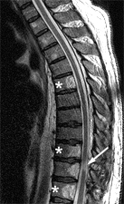
 Rare Case Of Multiple Aggressive Vertebral Hemangiomas Abstract Europe Pmc
 Vertebral Hemangioma Wikipedia

 Vertebral Hemangioma Radsource
 Atypical Mr Imaging Features Of Vertebral Hemangioma Involving The T12 Download Scientific Diagram